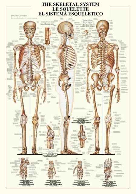
This instructional diagram presents the human skeletal system through three primary, full-body views: an anterior view on the left, a lateral profile view in the center, and a posterior view on the right. Each figure is rendered with high anatomical detail, showcasing the natural curves of the spine, the cage of the rib structure, and the complex articulation of the limbs and pelvis. The background is a clean, off-white parchment tone, providing a neutral field that keeps the focus entirely on the physiological structures.
Surrounding the central figures are several enlarged, detailed illustrations of specific bone joints and appendages, including close-ups of a knee joint and various views of the hands and feet. Each part of the skeletal anatomy is accompanied by lines pointing to specific features, which correspond to detailed labels written in English, French, and Spanish. The layout is symmetrical and balanced, with the title blocks centered at the top.
The illustration style is reminiscent of classic medical textbooks, utilizing a palette of bone-toned browns, tans, and deep shadows to provide a sense of depth and realism. The lighting is soft and consistent, emphasizing the texture and structural integrity of the bones. The overall tone is academic and clinical, designed for clear education and reference.